Implantation thoracoscopique

de pacemaker

Thoracoscopic vs conventional trans-diaphragmatic pacemaker implantation : comparative study in 4 cases

Auteurs: Drs. S.Libermann et É.Bomassi 24-04-2013
Centre Hospitalier Vétérinaire des Cordeliers, 29 avenue du Maréchal Joffre, 77100 Meaux.
E-mail: slibermann@chvcordeliers.comebomassi@chvcordeliers.com
Conférence

Introduction

Transvenous pacemaker implantation is safe

  • All dogs survived implantation in studies [1] and [2], 6 cardiac arrest in [3]
  • Major complications (15/104, Johnson and al. – 12/105, Wess and al., 51/136 Oyama and al.)
  • Lead displacement >>> infection, bleeding

Transvenous pacemaker implantation is successful in reducing clinical signs in sick sinus syndrome or third degree atrioventricular block

  • Mean survival times (2,2 years Wess and al.)
  • One-, three and five years survival : 86%, 65%, 39% (Johnson and al.)
  • One, two and three years survival : 70 %, 57 %, 45 % (Oyama and al.)

→ Gold Standard

[1] Results of pacemaker implantation in 104 dogs, M.S. Johnson, M.W.S Martin, W Henley, (2007) JSAP 48:4-11
[2] Applications, complications and outcomes of transvenous pacemaker implantation in 105 dogs (1997-2002), G Wess, W.P. Thomas, D.M. Berger, M.D. Kittleson, (2006) J Vet Intern Med 20(4):877-84
[3] Practices and outcomes of artificial cardiac pacing in 154 dogs, M.A. Oyama, D.D. Sisson, L.B. Lehmkuhl (2001), J Vet Intern Med 15(3):229-39

Interests of transvenous pacemaker implantation

  • Minimally invasive

Limits of transvenous pacemaker implantation :

  • Injury / sepsis in the area of right jugular vein
  • Price
  • Available
  • NO SURGEON NEEDED

Study design

Prospective case serie

Animals

  • Sick sinus syndrome (1), Sinoatrial block (1),
  • Atrioventricular block and non active or old myocarditis suspected (2)

Cadaveric study conducted to compare the epicardial access for implantation

Procedure

Dogs randomly assigned to:

- group 1 = conventional Trans-diaphragmatic approach(TDA)

- group 2 = Thoracoscopic implantation (TI)

  • Dorsal recumbancy
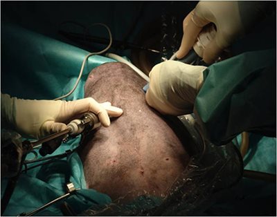
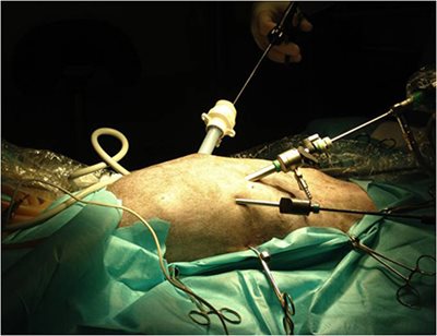
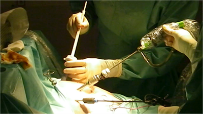
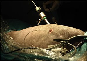
  • 5 mm telescope / paraxyphoïd approach
  • Instruments portals en in 6th intercostal space – left 15mm – Right 5 mm
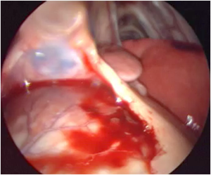
  • Pericardial window
  • Myocardic biopsy
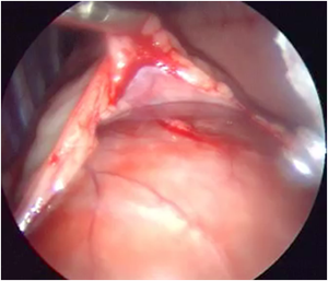
photo 1 photo 2

Photos 1&2: Thoracoscopic pericardectomy performed without pulmonary exclusion in 9 dogs.
Dupré G, Corlouer J, Bouvy B (2001). Vet Surg 30(1)21-7
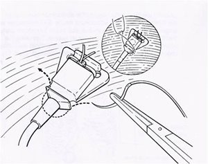
photo 3 schemaLeads

  • Medtronic Unipolar electrodes for single chamber pacing
  • Capsure EPI 4965 coated with steroïds
  • Suturelessunipolar myocardial screw-in pacing lead
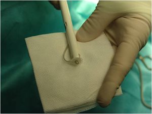
photo 4

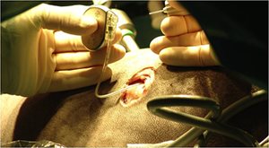
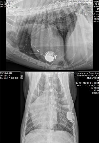
  • Pulse generator : VVIR Medtronic ADAPTA ADSR 01
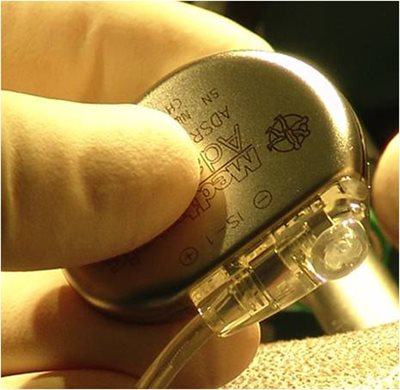
photo 5 : pulse generator photo 8

photo 6 photo 7
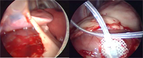
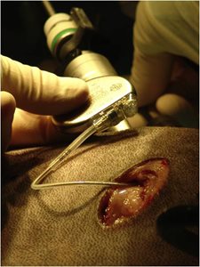
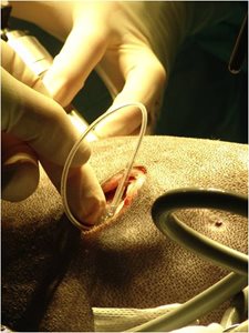
  • 15 mm portal for lead implatation
  • Lead and electrode positionning
  • Controle pacing
photo 9
photo 11 photo 12
photo 14 photo 13
photo 10

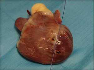
Anatomic study

  • Both procedures done on a cadaver
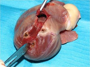
  • Epicardium marked on the insertion sites by Ligasure
  • Full heart dissection
photo 15 photo 16

Results

- Animals

 Group

Male
Female

Weight

Age

Breed

1 al 1

M

9,6 kg

11

WHWT

1 al 2

M

35 kg

7

Labrador

2 al 1

M

37 kg

10

S. Husky

2 al 2

F

27,5 kg

11

Weimaraner

- Surgical time

  • 46’ and 43 ‘ in Group 1
  • 44’ and 22’ in Group 2

- 1 major complication in Group 2 case 2

  • Pigtail loss, thoracoscopic reimplantation neededTwiddler syndrome

- Biopsy

  • Old myocarditis confirmed in both cases of Group 2
  • Biopsy had no consequence on cardiac pacing

- Outcomes

  • Hospital discharge 3 and 4 days PO in group 1 1 and 2 days PO in group 2
  • All dogs free of cardiac symptomes

Discussion

- Anatomic study

  • Transdiaphragmatic approach : Left ventricle
  • Thoracoscopic implantation : Right ventricle
  • +: more consistent with human guidelines
  • -: thinner myocardium in dogs  risk of pigtail loss??

- Right 6th intercostal portal can be used for electrode implantation

photo 17

- Feasability of thoracoscopic pacemaker lead implantation

  • Easy
  • Faster than TDA
  • Early hospital discharge

- Complication in group 2 (twiddler syndrome)

  • Lead displacement due to anormal coiling
    → wrong position of the generator
  • NOT due to the thoracoscopy
photo 18

- Thoracoscopic implantation > Thoracotomy

- TI > Trans-diaphragmatic approach

- T I > trans-xyphoïd approach ?

photo 19

Minimal invasive transxiphoie approach to the cardiac apex and caudoventral intrathoracic space, D.A. Nelson and al. (2012) Vet Surg 41:915-7

- Risks in endovascular implantation :

  • Thrombosis [1]
  • Obstruction [2,3]
  • Stricture [4]

- TI possible for dual chamber pacing : access allowed to the right auricule

[1]Cranial vena cava thrombosis associated with endocardial pacing leads in three dogs.
Murray J, O’Sullivan ML? Hawkes KC. J Am Anim Hosp Assoc. 2010 May-Jun;46(3):186-92
[2] Caudal vena cava obstruction caused by redundant pacemaker lead in a dog
Stauthammer C, Tobias A, France M, Olson J J Vet Cardiol 2009 Dec;11(2):141-(
[3] Cranial vena caval syndrome secondary to central venous obstruction associated with a
Pacemaker lead in a dog Mulz JM, Kraus MS, Thompson M, Flanders JA J Vet Cardiol 2010. Dec 12(3):217-23
[4] Succesful treatment of pacemaker-induced stricture and thrombosis of the cranial vena cava in two dogs by use of anticoagulants and balloon venoplasty. Cunningham SM, Ames MK, Rush JE, Rozanski EA.
J Am Vet Med Assoc 2009 Dec 15;235(12):1467-73

Conclusion

  • TI seems to be effective
  • Minimal invasive surgery
  • Can be an interesting alternative to endovascular implantation?
Retour

Informations pratiques

CHV des Cordeliers - Ouverture 24h/24 et 7j/7

Retour
  • Lun
    Consultations de 08h à 19h +Urgences 24h/24
  • Mar
    Consultations de 08h à 19h +Urgences 24h/24
  • Merc
    Consultations de 08h à 19h +Urgences 24h/24
  • Jeu
    Consultations de 08h à 19h +Urgences 24h/24
  • Vend
    Consultations de 08h à 19h +Urgences 24h/24
  • Sam
    Consultations de 08h à 19h +Urgences 24h/24
  • Dim
    Urgences 24h/24
Retour

Adresse de la clinique

35 Av. du Maréchal Joffre 77100 MEAUX
retrouvez-nous sur Google Maps